The identification features of the particular slide with little bit of explanation is given. The points in bold are specific features for a particular system and for a particular tissue.


Bone Transverse Section:


Hyaline cartilage:
- Covered by outer fibrous and inner chondrogenic perichondrium
- Homogeneous, glossy, transparent, basophilic matrix due to same refractive index for collagen type II fibers and ground substance
- Territorial matrix surrounding cell nest and interterritorial matrix between cell nests
- Chondrocytes arranged in the form of cell nest prevalent in the center
- Cell nest/ isogenous group- 2 to 4 cells present in groups surrounded by territorial matrix

Elastic cartilage:
- Covered by outer fibrous and inner chondrogenic perichondrium
- Matrix with more elastic fibers along with collagen type II fibers embedded in ground substance
- Larger chondrocytes in lacunae more prevalent in the center

White fibro cartilage:
- Devoid (absence) of perichondrium
- Collagen fibers running parallel to each other in bundles and less of ground substance in the matrix
- Chondrocytes arranged in rows

Bone:
In the same diagram both transverse section and longitudinal section are drawn.Bone Transverse Section:
- Covered by periosteum with outer fibrous and inner osteogenic layers
- Matrix with Haversian systems (Haversian canal surrounded by concentric lamellae)
- Outer circumferential lamellae present deep to periosteum, interstitial lamellae present between Haversian systems, inner circumferential lamellae covers endosteum
- Lamellae are thin plates of fibers and calcified matrix
- Lacunae are spaces with osteocytes present between two lamellae
- Canaliculi are thin spaces extending from lacunae with cytoplasmic processes
- Haversian canals run longitudinally connected by transverse channels called Volkmann's canal
- Lamellae arranged parallel to the Haversian canals
- Between adjacent lamellae are lacunae with osteocytes
- Haversian canals are surrounded by concentric lamellae, Volkmann's canals pass through lamellae connecting two Haversian canals. They contain blood vessels and nerves providing nutrition to bone

Skeletal muscle Transverse Section:
- Whole muscle surrounded by epimysium, bundle of muscle fibers (fascicle) by perimysium and single fiber by endomysium
- Nuclei are situated peripherally beneath sarcolemma
- Epimysium- dense irregular connective tissue, perimysium- less dense irregular connective tissue, endomysium- thin connective tissue

Skeletal muscle Longitudinal Section:
- Unbranched, long, voluntary cylindrical fibers with sarcolemma, sarcoplasm
- Multinuclei placed beneath sarcolemma in each muscle fiber
- Cross-striations- light I band and dark A band formed by contractile proteins actin and myosin
- Sarcolemma- cell membrane, plasma membrane
- Sarcoplasm- cytoplasm
- Sarcomere- functional contractile unit of muscle fiber having contractile proteins actin and myosin, extends between two Z lines
- I band is formed by only actin filaments attached to Z line. A band is formed by myosin filaments in the center and overlapped by actin filaments at the side


Cardiac muscle:
- Branched, short, involuntary cylindrical fibers
- Each muscle fiber has one or two nuclei in the center
- Cross-striations- light I band and dark A band formed by contractile proteins actin and myosin
- Intercalated disc- Cell junctional complex present between adjacent muscle fibers

Smooth muscle:
- Spindle shaped, non striated, involuntary muscle fibers
- Each muscle fiber has oval nucleus in the center
- It is found in the walls of digestive system, respiratory system, urinary system, genital system, blood vessels
- It appears smooth since the protein filaments are not arranged regularly in order

Peripheral nerve TS:
- Whole nerve is surrounded by connective tissue epineurium, fascicle by perineurium and each nerve fiber by endoneurium
- Between individual axons are seen numerous nuclei of Schwann cells
- Each axon is surrounded by myelin sheath
- Along with the connective tissue, there are blood vessels, nerves, lymphatics, adipose tissue

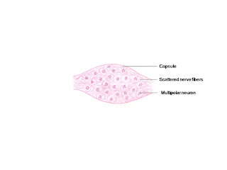
Sympathetic / autonomic ganglion:
- Multipolar, irregular shaped, smaller, scattered neurons
- Eccentric nucleus in each neuron
- Few satellite cells present around neurons
- Irregular shape of the cell is due to attachment of dendritic processes
- The satellite cells provide the structural and
- metabolic support to the neurons and insulate the neurons.
- Randomly arranged bundles of nerve fibers

Spinal ganglion/ Dorsal root ganglion:
- Pseudounipolar, larger neurons present in groups
- Fascicles of nerve fibers pass between unipolar neurons
- Nucleus with prominent nucleolus present in the center of neuron
- Satellite cells surrounding the neurons are more
- Fascicles of nerve fibers are the peripheral processes of bifurcating single axon of unipolar neuron
- The satellite cells provide the structural and metabolic support to the neurons and insulate the neurons.

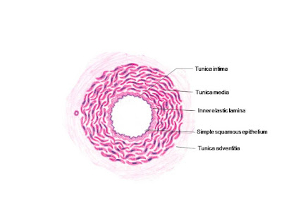
Large sized artery/ Elastic artery:
- Three layers from inside out- tunica intima, tunica media, tunica adventitia
- Tunica intima- lined by squamous cells (endothelium) surrounded by subendothelial connective tissue
- Tunica media is very thick with more number of elastic fibers, few smooth muscle fibers
- Tunica adventitia- connective tissue, vasa vasorum, nerves
- Subendothelial layer- loose connective tissue, few smooth muscle fibers, elastic fibers
- More number of elastic fibers give the artery the resilience where the artery can expand when more blood flows through the vessel and come back to its original position, so that there is steady flow of blood
- Example- aorta, brachiocephalic trunk, common carotid artery, subclavian artery

Medium sized artery/ distributing artery:
- Three layers from inside out- tunica intima, tunica media, tunica adventitia
- Tunica intima- lined by squamous cells (endothelium) surrounded by subendothelial connective tissue. Internal elastic lamina is more prominent
- Tunica media is as thick as tunica adventitia with more number of smooth muscle fibers.
- Tunica adventitia- very thin with connective tissue, vasa vasorum, nerves
- More number of smooth muscle fibers allows the artery to distribute blood to most parts of the body by its contraction

Medium sized vein:
- Three layers from inside out- tunica intima, tunica media, tunica adventitia
- Tunica intima- lined by squamous cells (endothelium) surrounded by subendothelial connective tissue.
- Tunica media - very thin.
- Tunica adventitia- very thick with connective tissue, vasa vasorum, nerves

Large sized vein:
- The thickness of the wall is half the size of the corresponding artery. Three layers from inside out- tunica intima, tunica media, tunica adventitia
- Tunica intima- lined by squamous cells (endothelium)
- Tunica media is thin with connective tissue and smooth muscle fibers
- Tunica adventitia is very thick compared to other two layers with connective tissue, vasa vasorum, nerves, longitudinally running smooth muscle fibers

Lymph node:
- Covered by connective tissue capsule sending incomplete trabeculae into lymphnode along with blood vessels and nerves
- Cortex: lymphatic nodules with germinal centers (B lymphocytes) , paracortex in deeper zone (T lymphocytes)
- Subcapsular sinus deep to capsule, trabecular sinus around trabeculae
- Medulla: medullary sinus, medullary cords
- Lymph flow: afferent lymphatic vessels-- subcapsular sinus--trabecular sinus--medullary sinus--efferent lymphatic vessel
- Sinuses- spaces with lymph supported by network of fine reticular fibers
- Lymph nodes filter the lymph by phagocytosis and acts as the principal site in which T and B lymphocytes undergo antigen-dependent proliferation and differentiation into effector lymphocytes (plasma cells and T cells) and memory B cells.
- Germinal center is the active site of lymphocyte proliferation

Thymus:
- Covered by connective tissue capsule sending incomplete trabeculae into thymus along with blood vessels and nerves.
- Each lobule is divided into cortex and medulla
- Cortex has dense aggregations of thymic lymphocytes
- Medulla has few lymphocytes, more epithelial reticular cells and Hassall's corpuscles
- Thymus shows age changes. Just after birth it is at its greatest development and by puberty it starts to regress and is replaced by adipose tissue
- Hassall's corpuscles- oval hyalinized structures with round aggregations of flattened epithelial cells and central calcified or degenerated epithelial cells
- Blood-thymus barrier- endothelial cells, epithelial reticular cells, macrophages which does not allow the substances transported in blood vessel to interact with developing T cells in the cortex

Spleen:
- Covered by connective tissue capsule sending incomplete trabeculae into spleen along with blood vessels and nerves.
- Divided into red pulp and white pulp
- Red pulp: Splenic cords of Billroth (small lymphocytes, red blood cells, neutrophils, eosinophils, ), pulp arteries and venous sinuses
- White pulp: lymphatic nodules with germinal centers and central artery

Tonsil:
- Free surface is covered by stratified squamous nonkeratinized epithelium which invaginates into tonsil to form tonsillar crypts
- The crypts are surrounded by numerous lymphatic nodules
- Attached surface is covered by connective tissue capsule which sends septa towards surface of tonsil. It acts as barrier from spreading tonsillar infections.
- Function of stratified squamous nonkeratinized epithelium is protection

Thick skin:
- Epidermis: 5 layers- stratum basale, stratum spinosum , stratum granulosum , stratum lucidum , stratum corneum
- Dermis: papillary layer and reticular layer with sweat glands
- Stratum basale- divides to increase number of cells, differentiate into other cells
- Stratum spinosum- polygonal cells, intermediate keratin filaments increase in number
- Stratum granulosum- intermediate keratin filaments and keratohyalin granules join to form keratin
- Stratum lucidum- lacks nucleus, densely packed keratin, contain eleidin
- Stratum corneum- lacks nucleus and organelles, full of keratin

Thin skin:
- Epidermis: 4 layers, stratum lucidum absent. Stratum basale, stratum spinosum, stratum granulosum, stratum corneum
- Dermis: papillary layer and reticular layer with hair follicle, sebaceous glands, arrector pili muscle and sweat glands
- Hair follicle- from inside out- inner root sheath, outer root sheath, connective tissue sheath
- Sweat glands- simple coiled tubular glands, lower coiled part forms secretory portion, upper tube forms duct, secrete non-viscous fluid
- Sebaceous gland- simple or branched acinar glands, open into hair follicle, secrete sebum
- Arrector pili muscle- smooth muscle attached to dermal papillae and hair shaft below the sebaceous gland, when it contract hair becomes straight

Serous salivary gland:
- Covered by connective tissue capsule which send septa along with blood vessels and nerves into gland dividing into lobes and lobules
- Lobe has numerous serous acini, numerous intercalated ducts with less connective tissue
- Salivary glands are compound tubuloacinar glands
- Acini or alveoli- small, sac-like dilations located at the end of the first part of the excretory duct system called the intercalated ducts
- Serous acini- serous cells surround small lumen. Each serous cell is pyramidal in shape with round nucleus in the lower half and zymogen granules in the upper half due to which cytoplasm stains dark with eosin
- Intercalated duct- simple cuboidal epithelium, striated duct- simple cuboidal to columnar epithelium with infoldings of the cell membrane at the base, interlobular/ interlobar duct- pseudostratified columnar to stratified cuboidal epithelium
- Myoepithelial cells contractile in nature, present in both type of alveoli, intercalated ducts. Flat, highly branching cells surround the acini between acinar cells and basement membrane. Helps in expelling secretory products from the acini.

Mucous salivary gland:
- Covered by connective tissue capsule which send septa along with blood vessels and nerves into gland dividing into lobes and lobules
- Lobe has predominantly mucous acini, few serous acini, few intercalated ducts, more of striated ducts
- Mucous acini- mucous cells surround large lumen. Each mucous cell is columnar in shape with flat nucleus pushed towards the basement membrane and the cytoplasm appears pale, frothy due to washing off of mucinogen granules during the slide preparation

Mixed salivary gland:
- Covered by connective tissue capsule which send septa along with blood vessels and nerves into gland dividing into lobes and lobules
- Lobe has mucous acini, serous acini, serous demilune (crescents of Gianuzzi), very few or absent intralobular ducts, more number of interlobular ducts, more amount of connective tissue
- Serous demilune / crescents of Gianuzzi- mucous acini capped with serous cells in the form of half moon or crescent shape

Placenta:
- It has fetal part and maternal part
- Fetal part: covered by amniotic membrane, mesoderm with blood vessels, chorion.
- Chorionic villi- Primary chorionic villi (core of cytotrophoblast cells covered by syncitiotrophoblast), secondary villi (core of mesoderm covered by cytotrophoblast and syncitiotrophoblast), tertiary villi (core of mesoderm with fetal blood vessels covered by cytotrophoblast and syncitiotrophoblast)
- Intervillous spaces: filled with maternal blood
- Maternal surface formed by decidua basalis, with endometrial blood vessels
Umbilical cord:
- Covered by amniotic membrane
- It has two umbilical arteries and one umbilical vein surrounded by Wharton's jelly (embryonic mesoderm)
- Wharton’s jelly is a mass of loose connective tissue with fibroblasts separated by fine collagen fibers and ground substance containing hydrated glycosaminoglycans rich in hyaluronic acid
- The two umbilical arteries carry deoxygenated blood from the fetus to the placenta and one umbilical vein that returns oxygenated blood from the placenta to the fetus.
Wonderful page.... Thanks a lot!!!
ReplyDeleteThank you 😊 😊 very useful info for medicozzz
ReplyDeleteKindly post diagram of medium sized vein also
ReplyDeleteThank u so much ..this information is very useful
ReplyDeleteSuperb! crisp and to the point. Very useful.
ReplyDeleteTaste bud diagram please
ReplyDeleteCan anyone share link for systemic histology?
ReplyDeleteclick on 'special histology- specific points' on the top right
DeleteCheck the blog archive under february 2016 post
DeleteHi, I writing to you from Azerbaijan. We are planning to print an album containing histology drawing, diagram for our students. Can we use yours?
ReplyDeleteThank you
ReplyDeletePlease use the diagrams. It should be of use to the students
Nice
ReplyDeleteI am Sherif Ibrahim a lecturer of Histology and cell biology and graduate of Michigan State University, USA. I see the great effort and awesome work you did in this blog and I Would like to cooperate with you. If you are interested please contact me on ibrahi22@msu.edu
ReplyDeleteThank you
Sherif Ibrahim
Thanks
ReplyDelete